The previous 5 MCQs on pedigrees have used dummy data that are good for teaching as they give nice clean and easily interpretable answers. However, the real world isn’t so clean! So, I have made a few more quizzes using data from published work to illustrate similar points with real examples.
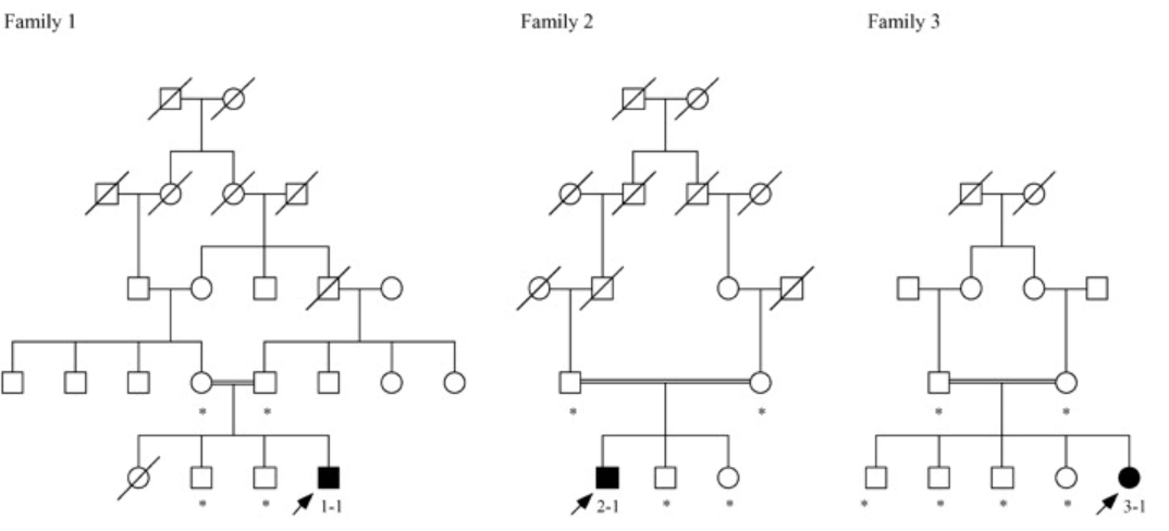
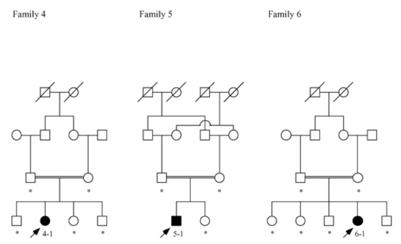
families 1-3families 4-6
The 6 pedigrees above all came from patients with a disease known as “laryngo-onycho-cutaneous syndrome” (LOCS). This is a rare disease where patients present with over production of granulation tissue in any of their exposed mucous membranes including eyes, throat and also have non-healing skin erosions. At the time this study began, the cause of the disorder was not yet known. However, a group of patients had presented all with very similar symptoms and all with distant relatives to the same part of the world.
Based only of the pedigrees, which of the following inheritance patterns are possible? There are more than one answer
Autosomal recessive
Autosomal dominant
Spontaneous mutation
Mitochondrial
X-linked recessive
X-linked dominant
Answer explanation
Click here to reveal the answer to the question above
Autosomal recessive – notice that in all the families, there are incidences of cousins marrying cousins (consanguineous marriages, indicated on the pedigrees by the double line connecting the parents). This doesn’t directly mean that there will be recessive inheritance, however, it does make it much more likely that recessive traits will present themselves. Spontaneous mutation – we can’t rule this out. There could be an easily mutated region that by chance has been mutated in these families. The consanguineous marriages made us think genetic but you can’t rule out chance! Wrong answers: autosomal dominant, X-linked dominant, one or more of the parents would be affected; X-linked recessive, in family 3, 4, 6 there are affected females being born to unaffected fathers. If X-recessive then the fathers would have a mutant X and present with the disease (as there are males with the disease, we know that there isn’t male suppression of phenotype or anything else a bit weird going on). Mitochondrial, no evidence of maternal transmission.
The DNA sequences above are from a region of a gene where (after further studies) there was a pathogenic mutation. The left trace is from an unrelated healthy person. The right trace is from one of the family members. Q1 what sort of mutation could have caused this trace?
heterozygous insertion or deletion
heterozygous splice site mutation
homozygous missense mutation
homozygous insertion or deletion
heterozygous missense mutation
homozygous splice site mutation
Answer explanation
Click here to reveal the answer to the question above
What you are seeing is two traces on top of one other, starting at the arrow. If you look carefully, you can see that there are now two green lines (As) beside each other in the mutant rather than one in the wild-type. This sort of sequence can be explained by a single base being inserted in one copy of this gene. Therefore it is a heterozygous insertion. It could be possible that two different insertions have occurred (e.g. a 2bp in one and 1bp in the other) but you can actually read the mutant sequence as two different letters based on the coloured traces and work out that it is just one mutant.
Q2 Which family members would a mutant trace like this come from? More than one answer is correct. Click all that apply
Father of patient 3-1
Mother of patient 3-1
Patient 3-1
Grandfather of patient 3-1
Grandmother of patient 3-1
Brother of patient 3-1
Answer explanation
Click here to reveal the explanation for the question above
Correct are either parent, the brother or either grandmother. wrong is grandfather of 3-1 or the patient 3-1 themselves. From the trace, we know that the person is heterozygous for the mutation. Therefore we can look at the pedigree for those likely to be het. The patient will be homozygous so not them. Parents must be het. Unaffected siblings could be wt (1/4) or het (1/2). The grandfathers married into the family and are unrelated to one another. It would be pretty unlucky for them to be the source. Much more likely is the related grandmothers carrying this rare mutation.
Q3 It turns out the mutation was 151insG – insertion of a G at nucleotide 151 – in the middle of the first exon of the LAMA3 gene. The whole LAMA3 gene is huge, several thousand base pairs and 72 exons. Giving a protein of ~1000 amino acids. What affect would one predict that this insertion mutation could cause?
No protein expression from the LAMA3 gene
Expression of a short protein of ~50 amino acids from the LAMA3 gene.
Expression of a mutant protein with the first 50 amino acids as normal and then 950 different amino acids.
Expression of the normal protein but at much reduced levels than normal
Expression of the mouse form of the protein instead of the human form
Answer explanation
Click here to reveal the explanation for the question above
We have a single base pair insertion into the first exon of a multi-exon gene. This will shift the reading frame of everything downstream of that mutation. The effect could be expression of a mutant protein. However, much more likely is that the shifted reading frame would introduce a stop codon at some early point. This could mean a short, ~50 amino acid protein (congrats if you said that). However, when you have a stop codon early in a transcript our cells recognise this as being a mistake and then target that mRNA for destruction in a process known as “nonsense mediated mRNA decay” (NMD). The result is that you would anticipate no protein to be made from the gene. Note that you may not have learned about this yet! NMD is a really important cell surveillance system to helps protect against deleterious proteins being produced.
Use the buttons below to try some of the other pedigree quizzes