
Part I – immunohistochemistry
As a little positive aside from all the COVID-19 related news, here is the first of a collection of cool science pics generated by the team over the last few months. Today’s post features a selection of immunohistochemistry images from human tissues.
Click on the first image to view as a slideshow.


What you are looking at:
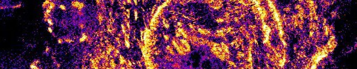
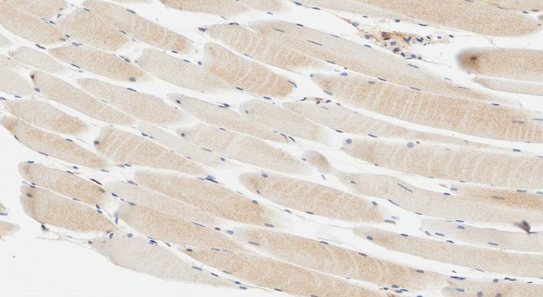
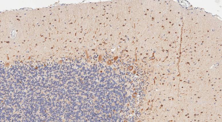
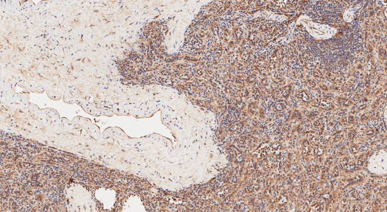
Each of these images is a thin slice from a block of tissue from a human donor. The tissue slice (section) has been probed with antibodies specific to a protein we are investigating. Those antibodies bind strongly to that protein but not to other parts of the tissue. We then use secondary antibodies which have an enzyme attached and probe the tissue section again. These secondary antibodies recognise the primary antibodies. The enzyme on the secondary antibodies are able to convert a substrate to produce the brown colour that you see in the images. This means we can interpret the image as meaning that where there is brown stain is where our protein of interest is located (and how brown roughly correlates with how much). The blue colour comes from a stain that binds to the cell nuclei, it’s useful to give contextual information.
Knowing where a protein is expressed is the first step to finding out its function. The 10 pics we have here are all analysing the same protein. These tissues are just some of a much larger panel that we analysed. In this case, no one has ever looked at this protein in these tissues before i.e. this stuff is totally new to the world. So they look cool (at least I think so), but the story they are telling is even cooler.
We would like your thoughts; comments below please. Also check back soon for part 2.
Also, have a look at our previous collections of cool fluorescence images here and TIRF images here

1 Comment