
I’m very pleased to tell you about our latest paper published this week in PLOS One.
Its been a long time coming, the manuscript co-first authored by two of the lab’s former PhD students Dr Lee Troughton, now at Loyola University and Dr Valentina Iorio now managing the teaching labs at University of Liverpool. Indeed, most of the team through the years contributed to this work with former PhD students and Dr Conor Sugden, Dr Liam Shaw, Dr Natasha Chavda, as well as last year’s Masters student Andrea Conway and our lab manager Dr Peter Wilson all pitching in important contributions.
It’s free to read/download here
So, what’s it about I hear you cry… well its about our favourite protein Laminin N terminus alpha 31 (click link for a friendly intro). This work builds on some of our previous studies. Specifically, in the past, we discovered that this protein is expressed in the cornea and that the expression changes during wound healing and stem cell activation (here), back during my PhD, when we found this protein, I showed that knockdown in skin cells affected the way they moved, and we’ve also determined that too much of the protein during embryogenesis changes the appearance of the formed skin (here), amongst other changes.
The new paper builds on all this work to finally (start) to understand how it is having these effects. We focused our work on corneal epithelium and used an overexpression system to mimic the changes we observed in tissues. Lee, Valentina et al then thoroughly characterised the cells, particularly honing in on the way that they attach.
There’s loads of stuff in there – the LaNt expressing cells are bigger and more tightly adhered, they assemble more mature hemidesmosomes, these two features are linked to changes in the way they deposit their laminin 332 and also how that laminin is also proteolytically processed (more than usual owing to increased activity of some of the most important matrix remodelling proteases MMPs). We partially establish causality by determining that we could reverse aspects of the phenotype by providing exogenous laminin or through blocking the activity of the MMPs, meaning the effects are matrix-mediated and MMP dependent.
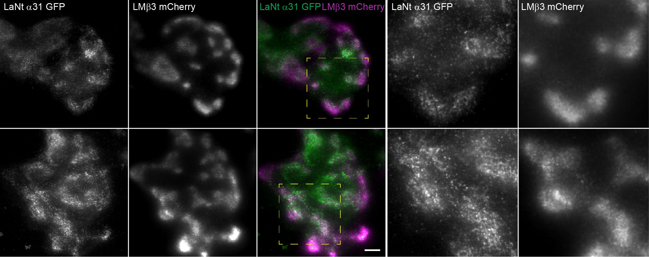
There’s also the first live imaging of LaNt dynamics relative to laminin b3. In the video LaNt a31 is imaged in the left panels (green in the merge to the right), Laminin b3 in the middle panels (magenta in merge) in corneal epithelial cells. We often talk about laminin patterns as “Cat paws”; our findings suggest that LaNt actually contributes to defining the patterning of the laminin…making it more cat paw-like!
Its cool.
Below is a still image using a slightly fancier imaging approach called total internal reflection, where you can see the co-distribution of the two proteins more easily.
Together, this paper delivers the first real understanding of how LaNt a31 works. It also creates more questions though. We’ve identified pieces of the puzzle but not yet found the links between those pieces.

Love the legacy on this one! So many people involved over so many years. Well done Kevin and Team! ________________________________
LikeLike