
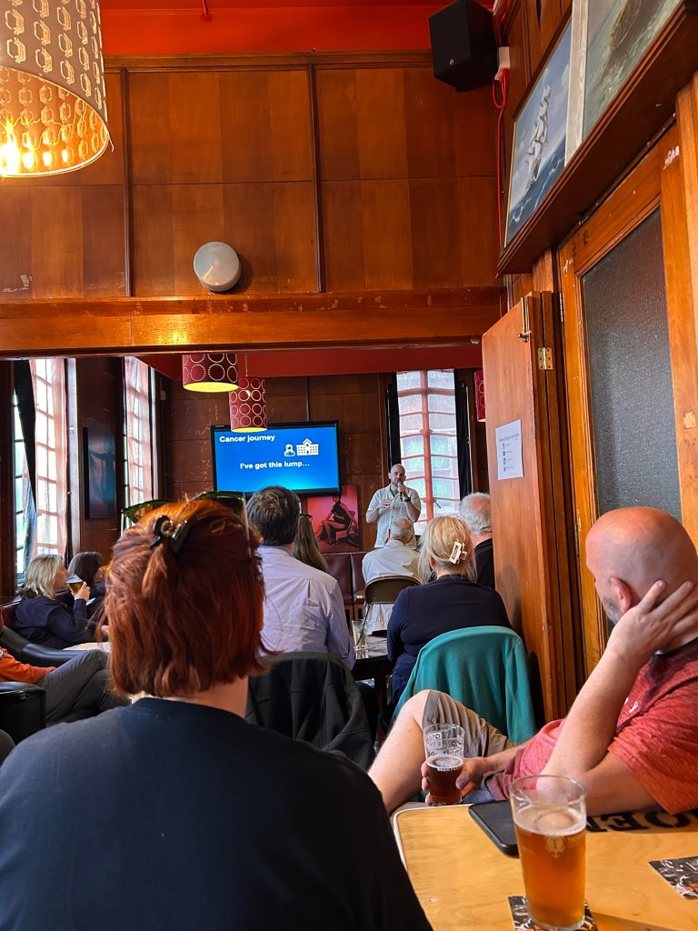
Last night, I had the privilege of delivering a talk at Pint of Science held at the Ship and Mitre in Liverpool. Pint of Science are fun events held internationally around this time of year. At each locally-organised event, scientific passion meets the warm, candid atmosphere of a local pub. My night was one of 11 sold-out events put on by University of Liverpool staff this week.
Public engagement is always fun. Mixing engagement with some nice beer makes it even better. Mixing it again with a chance to share some super cool imaging data makes it even more fun. I aimed for an intimate conversation about the wonders of microscopy and the secrets it unveils, particularly in the study of my favourite protein family, laminins. I think I ended up just enthusiastically telling the audience how cool everything was!

Laminins, as I am sure everyone reading this blog by now knows, are not just structural proteins; they are key functional protein of the extracellular matrix, orchestrating cell behaviour, tissue regeneration, and even influencing cancer progression. My talk was a deep dive into the microscope’s evolving ability to expose the hidden intricacies of these proteins. I took the audience on a visual journey: starting with the familiar terrain of conventional light microscopy that offers a panoramic view of tissue sections, moving into the detailed world of cell-level and live imaging, and finally, culminating in the awe-inspiring realm of super-resolution microscopy.
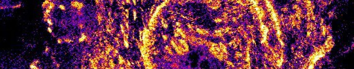
Really this was all just an elaborate set up to tell about our recent work using the iPALM microscope at Janelia—currently the best of its kind. This masterpiece of technology allows us to break the resolution barrier, capturing images with astonishing three-dimensional precision at the molecular level. It’s one thing to look at cells; it’s another entirely to witness the molecular substructure that was once hidden from view. The transition from conventional to super-resolution microscopy is, in a sense, like moving from a satellite image of a city to walking its streets and discovering the vibrant details of everyday life.

One of the most rewarding aspects of that night was not just the opportunity to share my passion for microscopy, but also to engage with a community of enthusiastic minds. Along with my own exploration of laminins, three other speakers presented on various facets of cancer research. Each talk, brimming with its own narrative and scientific insights, contributed to a mosaic of ideas that underscored how different types of research are contributing to our collective fight against complex diseases like cancer.

I found the conversations that followed the presentations to be as enlightening as the talks themselves. Science felt not habitual but alive—a dialogue between curiosity and evidence. Sharing these moments always reinforces why I continue to pursue the ever-deepening layers of my research. Every slide was a testament to the remarkable progress of my team and the field’s progress in advancing imaging technology. Every question from the audience ignited new sparks for future exploration.

In the end, last night wasn’t just about science—it was about the shared thrill of discovery and the promise of endless exploration. Together, as we peer deeper into the microscopic universe, we continue to unveil stories that are as transformative as they are beautiful. It was also about nice beer.

Read about our Super-res work in these blog posts – Janelia part 1, part 2, part 3, or see some cool images in these posts TIRF, Confocal 1, Confocal 2, histology

Great stuff!
LikeLike